This article is for general education and does not replace an in-person assessment, examination, or imaging. Everyone's injury pattern, medical history, and goals differ; use what you read here to prepare better questions for your doctor.
Dr. Nitin N Sunku is a consultant orthopedic and sports medicine surgeon. He sees patients at Raghava Multispeciality Hospital, Attibele, on Sarjapura–Attibele Road, and at Health Nest Hospital, HSR Layout, Bengaluru. If pain is rapidly worsening, you cannot bear weight, you develop numbness or weakness in a limb, or you have fever after an injury, seek urgent medical care. For non-emergency evaluation and individualised treatment options, book through the contact page.
Topics across this blog include knee ligament and meniscus problems, shoulder pain and instability, hip and knee arthritis, fracture recovery principles, spine symptoms when urgent causes have been excluded, running and tendon overuse issues, and what to expect from arthroscopy or joint replacement discussions. If you are comparing sources online, cross-check dates and always confirm advice with an in-person clinician.
Most people first hear the word "meniscus" the day it tears — after a twist on the football field, a deep squat gone wrong, or a knee that suddenly locks halfway through a flight of stairs. What often gets skipped in that moment is the why: why this small wedge of cartilage matters so much, why some tears heal quietly on their own while others never will, and why an MRI report can describe two tears that sound similar but carry completely different treatment plans.
Most people first hear the word "meniscus" the day it tears — after a twist on the football field, a deep squat gone wrong, or a knee that suddenly locks halfway through a flight of stairs. What often gets skipped in that moment is the why: why this small wedge of cartilage matters so much, why some tears heal quietly on their own while others never will, and why an MRI report can describe two tears that sound similar but carry completely different treatment plans.
This guide breaks down the basic science of meniscus tear — the anatomy, the tissue composition, the vascular zones, the biomechanics of load transmission, and the biological reasons behind every major treatment decision. Understanding this science does not just satisfy curiosity; it is what lets you ask the right questions before agreeing to surgery, physiotherapy, or a "wait and watch" plan.
What Is the Meniscus? A Quick Anatomical Refresher
Each knee contains two menisci — crescent-shaped, wedge-cross-section pads of fibrocartilage sitting between the femur (thigh bone) and the tibia (shin bone). The medial meniscus sits on the inner side of the knee and is C-shaped and comparatively fixed in place, which is part of why it is torn more often. The lateral meniscus sits on the outer side, is more O-shaped, and has greater mobility during knee movement, which paradoxically makes it less prone to tearing in everyday twisting injuries.
Both menisci attach to the tibia at their anterior and posterior "roots" and are stabilised further by the coronary ligaments, the transverse (intermeniscal) ligament, and — for the lateral meniscus — the meniscofemoral ligaments (of Humphry and Wrisberg). This attachment geometry matters clinically: a root tear effectively disconnects the meniscus from its anchor points and behaves, biomechanically, almost like removing the entire meniscus.
If you are trying to visualise the injury pattern rather than the underlying science, our detailed breakdown of medial meniscus tears and which activities to avoid during recovery is a useful companion read.
The Tissue Science: What the Meniscus Is Actually Made Of
At a cellular level, the meniscus is not simple cartilage — it is a specialised fibrocartilage built to handle both compression and tension simultaneously.
- Collagen composition: Roughly 75 percent of the meniscus's dry weight is collagen, and more than 90 percent of that is Type I collagen — the same tough, tension-resistant protein found in tendons. Smaller amounts of Type II, III, V and VI collagen are also present, contributing to the tissue's layered structure.
- Fibre orientation: Collagen fibres run in three directions — circumferential (the dominant pattern, running around the curve of the meniscus), radial (like tie-fibres holding the circumferential bundles together), and superficial mesh fibres near the surface. This circumferential orientation is the anatomical basis for "hoop stress," explained below.
- Cell type: The resident cells are called fibrochondrocytes, a hybrid between fibroblasts and chondrocytes, responsible for synthesising and maintaining this fibrocartilaginous matrix.
- Ground substance: Proteoglycans, glycoproteins and elastin fill the spaces between collagen bundles, giving the tissue its shock-absorbing, slightly compressible quality — similar in principle to how articular cartilage behaves, but structurally different and considerably tougher.
This composition is precisely why the meniscus behaves the way it does under load: strong enough to resist tension along its circumferential fibres, yet compressible enough to spread pressure evenly across the joint surface.
The Biomechanics: How the Meniscus Protects Your Knee
Understanding meniscus biomechanics explains almost every clinical decision that follows a tear.
Hoop Stress and Load Transmission
When your body weight compresses the femur down onto the tibia, the wedge-shaped meniscus is squeezed and wants to spread outward, like a doorstop being pushed further under a door. The circumferential collagen fibres resist this outward spreading, converting vertical compressive load into circumferential tension — a phenomenon called hoop stress. This mechanism allows the meniscus to distribute roughly 50–70 percent of the compressive load passing through the knee at 0 degrees of flexion, rising to as much as 85–90 percent at 90 degrees of flexion.
Load Sharing, Shock Absorption and Stability
Beyond hoop stress, the menisci perform several interlinked biomechanical jobs:
- Load sharing — increasing the contact area between the rounded femoral condyle and the relatively flat tibial plateau, which reduces peak stress on the articular cartilage underneath.
- Shock absorption — the viscoelastic matrix dampens impact loading during walking, running and jumping.
- Secondary stability — particularly the posterior horn of the medial meniscus, which acts as a secondary restraint to anterior tibial translation when the ACL is stretched or torn. This is exactly why ACL and meniscus injuries so frequently occur together.
- Joint lubrication and nutrition — helping distribute synovial fluid across the articular cartilage surfaces during motion.
A radial tear, a root tear, or a large meniscectomy disrupts this circumferential fibre continuity and effectively abolishes hoop-stress function in that segment — which is why even a "small" tear at the root can behave, mechanically, like losing the whole meniscus.
Why the Meniscus Tears: Traumatic vs Degenerative Pathophysiology
The basic science divides meniscus tears into two broad mechanistic categories, and this distinction drives almost every treatment conversation.
Traumatic (Acute) Tears
These occur when a rotational or shear force is applied to a loaded, partly bent knee — the classic "foot planted, body twists" mechanism seen in football, badminton, cricket fielding, or a deep squat under load. Acute tears are common in younger, more active patients and are frequently associated with ACL injury, since both structures resist the same rotational forces. Healthy, well-hydrated collagen in a younger meniscus tends to fail in vertical, longitudinal, or bucket-handle patterns.
Degenerative Tears
With age, the meniscus undergoes biochemical changes — reduced proteoglycan content, collagen fibre disorganisation, and microscopic mucoid degeneration within the substance of the tissue. This weakens the tissue's tensile strength long before any single traumatic event occurs, so degenerative tears can appear with minimal or no memorable injury — a deep squat while gardening, or simply standing up awkwardly. These tears are typically horizontal, complex, or flap-shaped and are frequently found alongside early osteoarthritis, since meniscal degeneration and cartilage degeneration usually progress together. Our detailed explainer on cartilage degeneration in the knee covers this overlapping process in plain language.
This is also why some tears are found incidentally on an MRI done for another reason, with no real symptoms at all — a pattern discussed further in our guide to asymptomatic meniscus tears.
Tear Patterns: Shape Determines Strategy
The basic science of meniscus tear classification is built around shape and orientation, because each pattern behaves differently under load and carries a different repair prognosis.
| Tear Pattern | Typical Cause | Repair Potential |
|---|---|---|
| Longitudinal/vertical | Traumatic, often young athletes | Good, especially if peripheral |
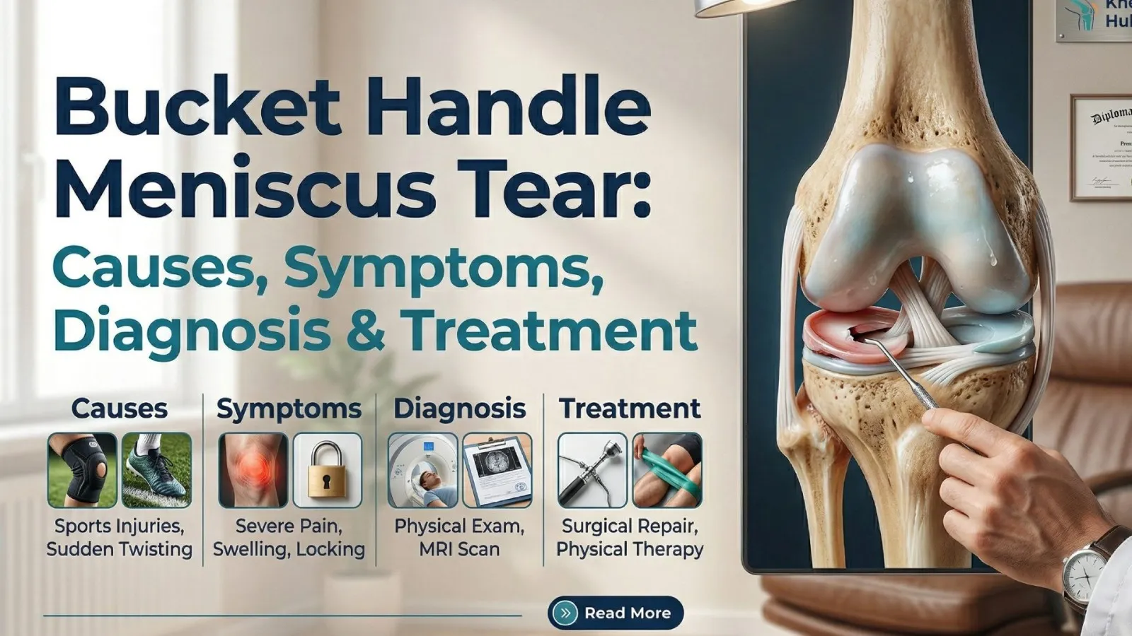
| Bucket-handle | Displaced longitudinal tear | Good if caught early; can cause locking |
| Radial | Traumatic or degenerative | Disrupts hoop stress severely; poor spontaneous healing |
| Horizontal | Degenerative | Often managed conservatively unless symptomatic |
| Flap (parrot-beak/oblique) | Degenerative or traumatic | Variable; mechanical symptoms common |
| Root tear | Traumatic or degenerative | Functions like total meniscus loss if untreated |
| Complex/degenerative | Chronic wear, aging | Usually poor repair candidate |
If you want a deep dive into a specific pattern, we have dedicated guides on the bucket-handle meniscus tear and the meniscus flap tear, both explaining diagnosis and recovery specific to that pattern.
The Vascular Zones: Why Some Tears Heal and Others Never Will
This is arguably the single most important piece of basic science for any patient trying to understand their treatment options. Blood supply to the meniscus enters from the outside in, via the perimeniscal capillary plexus fed by the geniculate arteries — and it does not reach the entire tissue.
- Red-red zone — the outer 10–30 percent of the meniscus, closest to the capsule. Fully vascularised. This is the zone with the highest healing potential, and tears here can sometimes heal on their own or respond very well to surgical repair.
- Red-white zone — the middle transition zone, with partial, reduced blood supply. Healing here is possible but less predictable, and outcomes depend heavily on tear size, stability, and the patient's age.
- White-white zone — the innermost portion of the meniscus, essentially avascular in adults and nourished only by diffusion from synovial fluid. Tears confined to this zone have very limited capacity for biological healing and are usually managed by removing the damaged, unstable fragment rather than attempting repair.
This zonal biology explains why two patients with a similarly sized tear can be given entirely different advice: one tear sits in a well-perfused red zone with genuine healing potential, while the other sits in a zone the body simply cannot repair on its own.
Meniscus, Cartilage and the Long-Term Osteoarthritis Link
Because the meniscus absorbs and redistributes so much load, damage to it — or its surgical removal — directly increases stress on the underlying articular cartilage. Multiple biomechanical studies confirm that meniscal injury and meniscal excision are among the strongest risk factors for accelerated cartilage wear and early-onset knee osteoarthritis. This is the central reason modern orthopedic practice has shifted decisively toward meniscus-preserving techniques — repair over removal wherever the tissue's vascularity and tear pattern make repair biologically realistic.
This is also why surgeons increasingly weigh long-term joint health, not just short-term symptom relief, when deciding between repair, partial meniscectomy, and non-surgical management. If arthritis has already progressed alongside the tear, our guide on knee osteoarthritis treatment explains how the two conditions are managed together.
How This Science Shapes Diagnosis
Because tear location, pattern, and vascularity all matter, imaging plays a central role. MRI remains the gold-standard, non-invasive method for evaluating meniscal lesions — it can usually indicate tear pattern, zone location, and stability with high sensitivity and specificity. Plain X-rays cannot visualise the meniscus directly but are still useful to rule out associated arthritis or bony injury. Ultrasound has limited value for typical meniscal tears, though it remains useful for guided injections elsewhere in the knee.
A careful clinical examination — assessing joint-line tenderness, effusion, locking, and provocative tests — is combined with imaging to reach a working diagnosis before any treatment path is discussed. If you are wondering whether your knee symptoms even warrant a specialist visit, this guide on when to see an orthopedic surgeon for knee pain may help you decide.
How the Science Translates Into Treatment Decisions
Every treatment recommendation for a meniscus tear ultimately traces back to the science above:
- Zone and vascularity decide whether repair is biologically feasible or whether the fragment needs to be trimmed.
- Tear pattern and stability decide whether the tear is likely to progress, catch, or displace if left alone.
- Patient age and activity level influence collagen quality, healing capacity, and functional demand on the knee.
- Associated injuries (ACL tear, early arthritis) change the urgency and the combined treatment plan.
This is why our approach to meniscus tear treatment always starts with correctly characterising the tear, rather than jumping straight to a generic "surgery or rest" answer. Depending on these factors, the plan may range from structured physiotherapy and activity modification to arthroscopic repair or partial meniscectomy, performed through the same keyhole techniques used in modern knee arthroscopy.
Key Takeaways
- The meniscus is a specialised, circumferentially-oriented fibrocartilage — not simple cartilage — built to convert compressive load into hoop-stress tension.
- Roughly 90 percent of its collagen is Type I, giving it tendon-like tensile strength combined with a compressible, shock-absorbing matrix.
- Blood supply only reaches the outer red-red zone; the inner white-white zone cannot heal on its own, which is the biological reason some tears are repaired and others are trimmed.
- Traumatic tears (young, active patients, twisting injury) and degenerative tears (older patients, gradual collagen breakdown) are mechanistically different and are managed differently.
- Preserving meniscal tissue whenever biologically realistic is now the guiding principle in orthopedic practice, because meniscal loss accelerates cartilage wear and osteoarthritis.
Frequently Asked Questions
What is the basic science behind a meniscus tear?
The basic science centres on the meniscus's fibrocartilage structure — mostly circumferentially-arranged Type I collagen — which converts knee compression into hoop-stress tension. A tear disrupts this fibre continuity, and how well it heals depends on which vascular zone (red-red, red-white, or white-white) it occurs in.
Why do some meniscus tears heal without surgery and others don't?
Healing potential depends almost entirely on blood supply. Tears in the outer, vascularised red-red zone receive nutrients and healing factors directly and can sometimes heal with rest, bracing, and physiotherapy. Tears in the inner, avascular white-white zone lack this blood supply and generally cannot heal biologically on their own, which is why they are more often treated by trimming the damaged fragment.
Is the meniscus cartilage or a ligament?
Neither, strictly speaking. The meniscus is a specialised fibrocartilage — structurally distinct from both the smooth hyaline (articular) cartilage lining the joint surfaces and from ligaments, which are dense, purely tension-resistant tissue. It combines features of both to handle compressive and tensile loads simultaneously.
What is the difference between a traumatic and a degenerative meniscus tear?
A traumatic tear results from an acute twisting or shearing injury to a normal, healthy meniscus, typically in younger or more active people. A degenerative tear develops gradually as collagen and proteoglycan content in the tissue break down with age, often appearing with little or no specific injury and frequently alongside early knee osteoarthritis.
Why is the meniscus important for preventing arthritis?
The meniscus distributes a large share of the compressive load passing through the knee and protects the articular cartilage from concentrated stress. When the meniscus is torn, degenerated, or removed, that protective load-sharing function is lost, and the underlying cartilage wears down faster — a well-documented pathway toward earlier knee osteoarthritis.
Can a meniscus regenerate after being torn?
Only within limits. The vascularised outer third has genuine, if modest, regenerative capacity, aided by fibrin clot formation and local growth factors. The avascular inner two-thirds have essentially no capacity for true tissue regeneration on their own, which is why researchers continue to explore biological augmentation techniques such as scaffolds and growth-factor therapies for these zones.
Does age affect meniscus healing?
Yes. Younger patients typically have better-hydrated, more organised collagen and a more robust peripheral blood supply, giving traumatic tears a higher chance of successful repair. With age, collagen disorganisation and reduced vascularity lower the baseline healing capacity, which is one reason degenerative tears in older adults are more often managed with activity modification and targeted rehabilitation rather than repair surgery.
This article is for general educational purposes and does not replace an in-person orthopedic evaluation. If you have persistent knee pain, swelling, locking, or a suspected meniscus tear, book a consultation with Dr. Nitin N Sunku for an accurate, examination-based diagnosis and treatment plan.
Further reading (external, evidence-based source): The Meniscus: Basic Science and Therapeutic Approaches — PMC, National Library of Medicine.
About the Author
Dr. Nitin N Sunku
MBBS, MS (Orthopedics), Fellowship in Arthroscopy & Sports Medicine
Dr. Nitin N Sunku is a Consultant Orthopedic & Sports Medicine Surgeon with over 10 years of focused practice in Bengaluru. He serves as the Team Doctor for Bengaluru FC and consults at Raghava Multispeciality Hospital (Attibele) and Health Nest Hospital (HSR Layout). His clinical interests include arthroscopy, ligament & meniscus care, regenerative orthopedic medicine, ultrasound-guided injections, and joint replacement.
Experiencing symptoms?
Don't let pain hold you back. Schedule a comprehensive evaluation with Dr. Nitin today.
Book Appointment