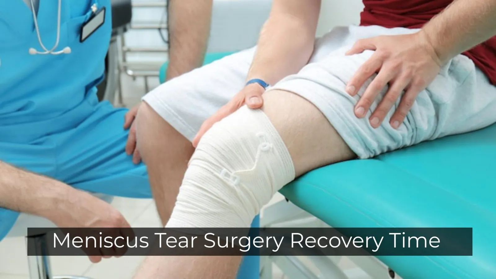
This article is for general education and does not replace an in-person assessment, examination, or imaging. Everyone's injury pattern, medical history, and goals differ; use what you read here to prepare better questions for your doctor.
Dr. Nitin N Sunku is a consultant orthopedic and sports medicine surgeon. He sees patients at Raghava Multispeciality Hospital, Attibele, on Sarjapura–Attibele Road, and at Health Nest Hospital, HSR Layout, Bengaluru. If pain is rapidly worsening, you cannot bear weight, you develop numbness or weakness in a limb, or you have fever after an injury, seek urgent medical care. For non-emergency evaluation and individualised treatment options, book through the contact page.
Topics across this blog include knee ligament and meniscus problems, shoulder pain and instability, hip and knee arthritis, fracture recovery principles, spine symptoms when urgent causes have been excluded, running and tendon overuse issues, and what to expect from arthroscopy or joint replacement discussions. If you are comparing sources online, cross-check dates and always confirm advice with an in-person clinician.
Outer knee pain after a twist or after ACL surgery? A specialist guide to anterolateral meniscus tears, root avulsions, diagnosis pitfalls, and modern arthroscopic root repair in Bengaluru.
A sudden twist during a football match, a deep squat at the gym, or an awkward landing from a jump. These are the moments that can cause an anterolateral meniscus tear or, in more serious cases, an anterior lateral meniscus root tear — two closely related knee injuries that are often misdiagnosed, under-reported, and misunderstood by patients and even some general practitioners.
The Lateral Meniscus and Its Root Attachments
The knee contains two C-shaped fibrocartilaginous structures called menisci. The lateral meniscus sits on the outer side of the knee, between the femur and tibia. It covers a larger portion of the tibial surface than the medial meniscus and plays a critical role in load distribution, shock absorption, and joint stability.
Each meniscus has an anterior horn and a posterior horn. At both ends, strong ligament-like root attachments anchor the meniscus securely to the tibial plateau. These roots allow the meniscus to function as a complete ring under compression, transmitting 50–70% of the body's weight across the tibiofemoral joint. When the anterior root of the lateral meniscus is torn, the meniscus loses its ability to distribute load — the result closely mimics what happens when the entire lateral meniscus is surgically removed, which accelerates joint deterioration and early-onset osteoarthritis.
Anterolateral Tear vs Anterior Lateral Root Tear: What's the Difference?
Anterolateral Meniscus Tear
A tear in the anterolateral zone of the lateral meniscus — the front and outer portion. Patterns can be horizontal, longitudinal, radial, or complex. Common after a direct blow to the outer knee, a rotational injury, or alongside an ACL rupture. Symptoms: outer knee pain, joint line tenderness, swelling, clicking or catching.
Anterior Lateral Meniscus Root Tear
A specific subtype involving the insertion point of the anterior horn into the tibial bone. The root sits in front of the intercondylar eminence, just beneath the footprint of the ACL tibial tunnel. Research shows that during ACL reconstruction, inadvertent damage to the anterior lateral meniscus root can occur in up to 18% of cases, particularly in patients with smaller anatomical structures. A complete root tear effectively converts the lateral meniscus into a functionally open ring — dramatically increasing tibiofemoral contact pressures.
Common Causes and Risk Factors
Acute causes
- ACL rupture with simultaneous rotational injury (the most common cause in young, active individuals).
- High-energy tibial plateau fractures.
- Sudden deep squatting or pivoting under load.
- Contact sports injuries — football, basketball, kabaddi, wrestling.
- Landing awkwardly from a jump or a misstep on uneven ground.
- Iatrogenic injury during ACL reconstruction (tunnel drilling near the anterior root footprint).
Degenerative and chronic causes
- Repeated high-flexion postures common in Indian daily activities — sitting cross-legged, squatting, Indian-style toilets.
- Age-related fibrocartilage degeneration over 40.
- Chronic ACL-deficient knee left untreated for years.
- Occupations requiring prolonged kneeling or crouching.
Symptoms You Should Not Ignore
- Outer (lateral) knee pain that worsens with weight-bearing, squatting, or twisting.
- Swelling along the outer joint line within 24–48 hours of injury.
- Clicking, popping, or catching when bending or straightening the knee.
- A feeling of the knee "giving way" during change-of-direction movements.
- Stiffness when trying to fully straighten or deeply bend the knee.
- Pain reproduced when pressing along the lateral joint line.
- In root tears, palpable extrusion of the meniscus at the outer joint margin.
Not every click or outer knee pain is a meniscus tear. A proper clinical evaluation by a sports medicine specialist is essential.
How It's Diagnosed
Clinical examination
- McMurray test — rotating the tibia while flexing the knee to reproduce clicking or pain.
- Thessaly test — weight-bearing on a single leg while rotating the knee at 20° flexion.
- Joint line tenderness palpation along the lateral joint line.
- Dial test and pivot shift assessment when ACL injury is also suspected.
MRI imaging — the gold standard
An anterior lateral meniscus root tear typically appears on MRI as a full-thickness signal disruption within 9 mm of the central root attachment. Key signs include the "ghost sign" (absence of the root on expected sagittal slices), meniscal extrusion on coronal views, and subchondral bone oedema.
Diagnostic arthroscopy
Where MRI is inconclusive or the clinical picture does not match imaging, diagnostic arthroscopy allows direct visualisation of the meniscus and root — particularly useful when planning surgical repair.
Classification of Root Tears
- Type 1: stable radial tear within 9 mm of root centre, partial attachment intact.
- Type 2: complete radial tear at or within the root zone.
- Type 3: bucket-handle tear with a root component.
- Type 4: complex degenerative tear involving the root.
- Type 5: bony avulsion of the root from the tibial attachment.
Treatment Options
Non-surgical management
Stable, partial tears with good tissue quality — particularly in the peripheral vascular zone — can sometimes be managed without surgery. Options include activity modification, targeted physiotherapy, anti-inflammatory medication, bracing, and selected corticosteroid or hyaluronic acid injections. However, complete root tears causing meniscal extrusion are unlikely to heal on their own.
Arthroscopic root repair
The gold-standard surgical technique for complete anterior lateral meniscus root tears. The goal is to anatomically reattach the avulsed root back to its native tibial footprint, restoring meniscal biomechanics and protecting the articular cartilage.
- Transtibial reinsertion: sutures pass through the detached root and a bone tunnel drilled in the tibia, then tied over a button or cortical fixation. Biomechanically the most effective at restoring tibiofemoral contact area.
- Suture anchor repair: for anterior horn tears (not root avulsions), suture anchors placed at the anterolateral edge of the tibial plateau secure the horn directly to bone.
- All-inside PCL fixation technique: for posterior lateral root tears coexisting with anterior pathology in complex combined injuries.
Partial meniscectomy — a last resort
When root or anterior horn tissue is severely degenerated or fragmented, partial meniscectomy may be necessary. While it relieves mechanical symptoms, it does not restore normal biomechanics. For young patients especially, every effort is made to preserve meniscal tissue before considering removal.
Recovery Timeline After Root Repair
Phase 1: Weeks 0–6 — protection
- Non-weight-bearing or partial weight-bearing with crutches for 4–6 weeks.
- Knee brace restricting range of motion (typically 0–90°).
- Gentle quadriceps activation, straight leg raises, ankle pumps.
- Swelling management with ice, elevation, and compression.
Phase 2: Weeks 6–12 — progressive loading
- Gradual return to full weight-bearing.
- Progressive range of motion to full flexion.
- Closed-chain strengthening — mini squats, leg press, step-ups.
- Pool-based rehabilitation to reduce joint stress.
Phase 3: Months 3–6 — functional rehabilitation
- Sport-specific agility drills, running, change-of-direction training.
- Balance and proprioception work to restore joint coordination.
- Return-to-sport criteria: symmetrical strength, no swelling, no pain.
Full return to competitive sport typically occurs between 4 and 6 months after surgery, depending on the sport and the individual.
What Happens If a Root Tear Is Left Untreated?
An untreated complete root tear causes the meniscus to extrude outward under body weight rather than distributing load evenly. Animal studies show early degenerative changes in tibiofemoral cartilage within months. In humans, the risk of tibiofemoral osteoarthritis rises measurably over 3–5 years. In younger patients, this accelerated degeneration can mean the difference between a career cut short and many more years of active sport. In middle-aged patients, it may push the knee replacement timeline significantly earlier than would otherwise be expected.
When Should You See a Doctor?
- Outer knee pain after a sports injury or twisting movement that does not improve within 1–2 weeks.
- An MRI report mentioning anterior horn lateral meniscus tear, root tear, meniscal extrusion, or ghost sign.
- Persistent clicking, locking, or giving way of the knee.
- Knee pain that recurs after ACL surgery on the same side.
- Progressive outer knee pain in an active adult over 40 without a clear history of injury.
Frequently Asked Questions
What exactly is an anterior lateral meniscus root tear?
A disruption of the ligamentous attachment that anchors the front portion of the lateral meniscus to the tibia. The meniscus loses its ability to function as a load-distributing ring, increasing pressure on the knee cartilage and the risk of early osteoarthritis.
How is this different from a regular meniscus tear?
A regular tear refers to any disruption within the body of the meniscal tissue. A root tear specifically disrupts the bony anchor of the meniscus — compromising the entire structural integrity of the cartilage.
Can it heal without surgery?
Partial or minor anterior horn tears in the vascular zone may heal with conservative management. Complete root tears rarely heal on their own — surgical root reinsertion is generally recommended for active individuals.
What is the recovery time after root repair?
Most patients are non-weight-bearing for 4–6 weeks, followed by progressive physiotherapy over 3–6 months. Return to competitive sport is typically 4–6 months.
Is a root tear commonly missed on MRI?
Yes. The ghost sign, meniscal extrusion of 3 mm or more, and subchondral bone oedema should raise suspicion. An experienced reader is essential.
Can a root tear occur during ACL surgery?
Yes. The anterior lateral meniscus root sits very close to the ACL tibial tunnel. Iatrogenic root damage is documented in up to 18% of ACL reconstructions in some studies, particularly in patients with smaller skeletal anatomy.
What are the long-term risks if untreated?
Progressive tibiofemoral cartilage damage, meniscal extrusion, and early-onset knee osteoarthritis. The risk of requiring knee replacement is meaningfully increased — especially in younger, active patients.
Educational content. If you have been diagnosed with or suspect an anterolateral meniscus tear or anterior lateral meniscus root tear, seek a specialist evaluation. Book a consultation at Health Nest Hospital, HSR Layout or Raghava Multispeciality Hospital, Attibele.
About the Author
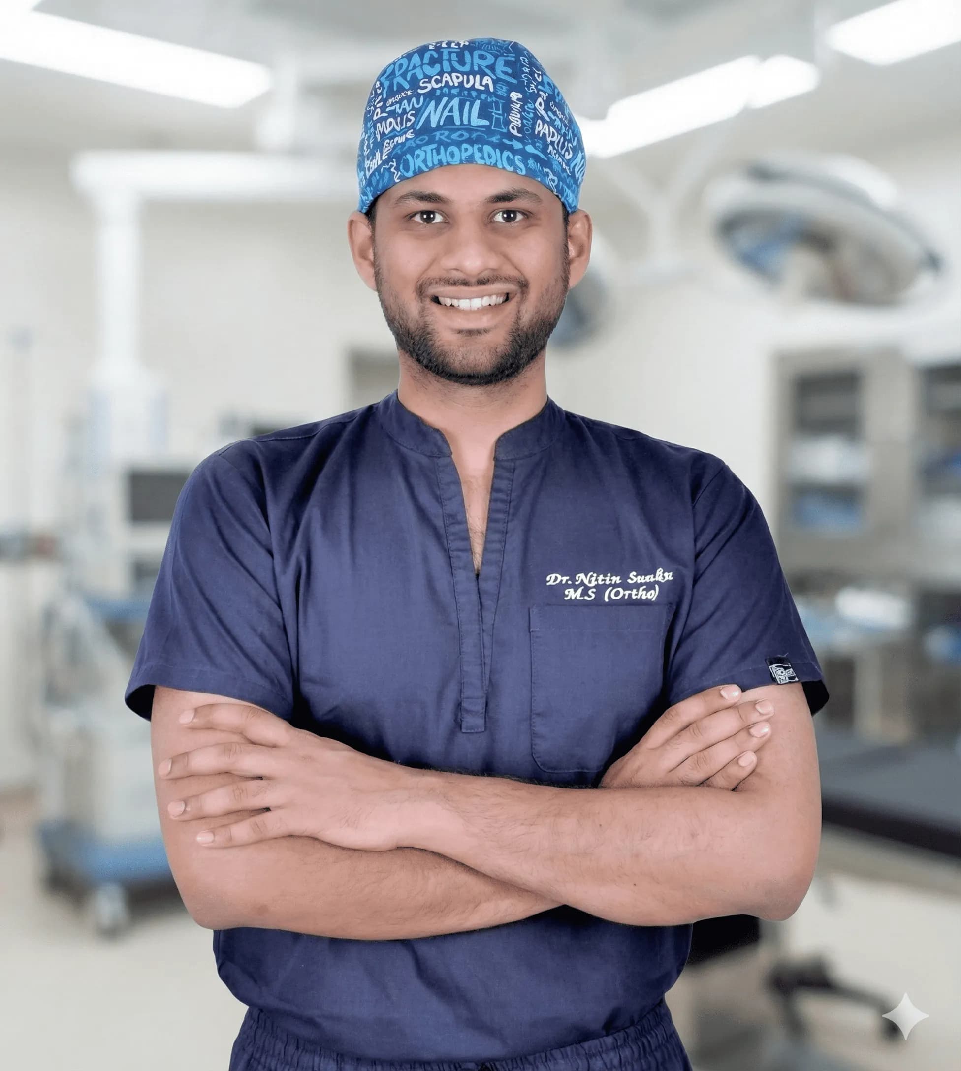
Dr. Nitin N Sunku
MBBS, MS (Orthopedics), Fellowship in Arthroscopy & Sports Medicine
Dr. Nitin N Sunku is a Consultant Orthopedic & Sports Medicine Surgeon with over 10 years of focused practice in Bengaluru. He serves as the Team Doctor for Bengaluru FC and consults at Raghava Multispeciality Hospital (Attibele) and Health Nest Hospital (HSR Layout). His clinical interests include arthroscopy, ligament & meniscus care, regenerative orthopedic medicine, ultrasound-guided injections, and joint replacement.
Experiencing symptoms?
Don't let pain hold you back. Schedule a comprehensive evaluation with Dr. Nitin today.
Book Appointment