This article is for general education and does not replace an in-person assessment, examination, or imaging. Everyone's injury pattern, medical history, and goals differ; use what you read here to prepare better questions for your doctor.
Dr. Nitin N Sunku is a consultant orthopedic and sports medicine surgeon. He sees patients at Raghava Multispeciality Hospital, Attibele, on Sarjapura–Attibele Road, and at Health Nest Hospital, HSR Layout, Bengaluru. If pain is rapidly worsening, you cannot bear weight, you develop numbness or weakness in a limb, or you have fever after an injury, seek urgent medical care. For non-emergency evaluation and individualised treatment options, book through the contact page.
Topics across this blog include knee ligament and meniscus problems, shoulder pain and instability, hip and knee arthritis, fracture recovery principles, spine symptoms when urgent causes have been excluded, running and tendon overuse issues, and what to expect from arthroscopy or joint replacement discussions. If you are comparing sources online, cross-check dates and always confirm advice with an in-person clinician.
A complete guide to anterior cruciate ligament anatomy — origin, insertion, the two functional bundles, and anterior and posterior cruciate ligament function compared.
When a patient walks into our clinic in Bengaluru with knee pain after a football collision or a sudden pivot on the basketball court, one of the first structures we evaluate is the anterior cruciate ligament (ACL). Most patients have heard of an ACL tear but have little understanding of anterior cruciate ligament anatomy — where this ligament actually sits, where it begins, and where it ends inside the knee. That knowledge gap matters clinically — because the ACL's origin and insertion directly determine how surgeons place bone tunnels during reconstruction, which graft angle restores natural mechanics, and how physiotherapists design loading protocols.
What Is the ACL?
The ACL is one of four main stabilising ligaments of the knee joint. It sits inside the joint capsule, within the intercondylar notch — the hollow space between the two rounded ends of the femur. The word cruciate comes from the Latin crux, meaning cross — because the ACL and its partner, the PCL, cross each other when viewed from the front.
The ACL is roughly 3–4 cm long and about 10 mm wide at its midpoint. It is composed primarily of Type I collagen fibres arranged in slightly twisted bundles — a design that distributes load across the ligament at different knee angles.
ACL Origin and Insertion: Exact Anatomical Points
Femoral Origin (where the ACL begins)
The ACL originates from the posteromedial surface of the lateral femoral condyle, inside the intercondylar notch — the inner wall of the outer bony knob at the end of the femur, positioned toward the back of the notch and slightly oval in shape.
- Sits posterior to the centre of rotation of the knee joint.
- Footprint is oval, approximately 18 mm long and 11 mm wide.
- The resident's ridge — a bony prominence along the condyle — marks the anterior border of the footprint and is a key intraoperative landmark.
Tibial Insertion (where the ACL ends)
The ACL inserts into the anterior intercondylar area of the tibial plateau, anterolaterally to the medial tibial eminence.
- Centre of the tibial insertion lies roughly 7–8 mm anterior to the PCL.
- Footprint spans approximately 13–16 mm long and 10–11 mm wide.
- A portion of the lateral meniscus anterior horn attachment overlaps the posterior edge of the ACL tibial footprint.
- The insertion area is larger than the femoral footprint, giving the ACL a slightly fan-shaped profile at the tibia.
Understanding these exact measurements is why ACL reconstruction at our clinic begins with careful preoperative planning using MRI to measure each patient's individual insertion dimensions before a single incision is made.
Course of the ACL Through the Knee
From its tibial insertion, the ACL runs superiorly, posteriorly, and laterally to reach the femoral condyle. As it travels through the intercondylar notch, it passes anterior to the PCL, and the two ligaments cross in an X pattern — hence the cruciate name.
The ligament is intra-articular (inside the joint) but extrasynovial (outside the synovial membrane). A synovial fold wraps around it, providing blood supply from branches of the middle genicular artery. This vascular envelope is thin, which partly explains why a complete ACL tear has a limited capacity to heal on its own.
The Two Functional Bundles
1. Anteromedial (AM) Bundle
- Originates from the proximal and anterior portion of the femoral footprint; inserts into the anteromedial aspect of the tibial footprint.
- Becomes taut as the knee flexes beyond 30°.
- Primary restraint against anterior tibial translation.
- Remains moderately lax in full extension.
2. Posterolateral (PL) Bundle
- Originates from the distal and posterior portion of the femoral footprint; inserts into the posterolateral part of the tibial footprint.
- Taut in full extension or near extension.
- Key contributor to rotational stability, particularly resisting internal tibial rotation.
- Becomes lax beyond 30–40° flexion.
The interplay between these bundles is why the pivot-shift test — which stresses the knee in slight flexion — provokes instability in ACL-deficient knees even when the anterior drawer test is equivocal.
Biomechanical Functions
- Resisting anterior tibial translation: prevents the tibia from sliding forward on a stationary femur.
- Controlling internal tibial rotation: especially in the 0–30° flexion range.
- Limiting knee hyperextension: secondary check when the knee is forced beyond straight.
- Proprioceptive feedback: mechanoreceptors sense joint position and velocity — a function partially lost after injury that must be retrained.
Because the femoral origin sits posterior to the knee's centre of rotation, the ACL becomes taut as the knee extends. This is the anatomical reason why straightening the knee (a blocked tackle, unexpected deceleration) combined with a rotational force creates the highest risk of ACL rupture.
Anterior and posterior cruciate ligament function
To understand anterior and posterior cruciate ligament function, it helps to look at them as a pair. The ACL and PCL both sit inside the intercondylar notch and cross each other in an X shape — that is where the name cruciate (cross) comes from. The ACL runs from the back of the lateral femoral condyle forward and downward to the front of the tibial plateau. The PCL runs from the inner wall of the medial femoral condyle backward and downward to the back of the tibia.
Functionally, the two ligaments are opposites that work together to keep the knee stable. The ACL primarily resists anterior tibial translation — it stops the tibia from sliding forward on the femur — and contributes to rotational stability. The PCL primarily resists posterior tibial translation — it stops the tibia from sliding backward. The PCL is also the thicker and stronger of the two, which is one reason isolated PCL tears are less common than ACL tears.
Injury mechanisms differ accordingly. ACL tears typically happen with sudden deceleration, pivoting, cutting, or awkward landings — the classic non-contact sports mechanism. PCL tears usually need a posteriorly directed force on the front of the tibia — the “dashboard injury” from a road traffic accident, or a fall onto a bent knee. Recognising which ligament has failed shapes the entire treatment plan, from bracing angle to surgical technique.
Why Origin and Insertion Matter for Reconstruction
For patients considering surgery, the accuracy of bone tunnel placement — guided by knowledge of the ACL's true origin and insertion — is the single most important surgical variable. Misplaced tunnels, even by a few millimetres, lead to abnormal knee mechanics, higher re-tear rates, and accelerated cartilage wear.
Anatomic vs Non-Anatomic Reconstruction
Older transtibial techniques often placed the femoral tunnel too anteriorly, producing a vertical graft that restored anterior translation reasonably well but failed to control rotation. Patients continued to report a give-way feeling during pivoting. Modern anatomic ACL reconstruction — the technique used in our practice — places each tunnel at the centre of the native ACL footprint via an independent anteromedial portal. The result is a more oblique graft that replicates the native ligament's orientation and restores both translational and rotational stability.
Single-Bundle vs Double-Bundle
Double-bundle reconstruction places two grafts to reconstruct both AM and PL bundles independently. It requires sufficient footprint space and is technically more demanding. For most patients — especially those with a tibial insertion width under 14 mm — a well-placed single-bundle anatomic reconstruction at the centre of the footprint achieves excellent outcomes.
Connecting Anatomy to Injury Mechanism
Common ACL injury mechanisms in our Bengaluru patient population:
- Sudden deceleration with a planting and cutting motion (football, kabaddi, basketball).
- Landing from a jump with the knee slightly bent and internally rotated.
- Direct contact causing forced valgus and rotation at the knee.
- Hyperextension — kicking and missing, or landing awkwardly.
Symptoms and Diagnosis
- A popping sound or sensation at the time of injury.
- Rapid knee swelling within hours (haemarthrosis).
- Feeling that the knee gave way or buckled.
- Inability to continue the sport or activity.
- Persistent instability during walking, turning, or stair use.
The Lachman test (anterior tibial translation at 30° flexion) and the pivot-shift test (rotational instability) are the most reliable bedside indicators. MRI confirms the diagnosis and identifies concurrent injuries — meniscus tears, the classic bone bruise pattern at the lateral femoral condyle and posterior lateral tibia, and chondral damage.
Treatment Options
Non-operative
A structured physiotherapy programme focused on quadriceps and hamstring strengthening, proprioception retraining, and activity modification can allow older, less active patients to live comfortably without an intact ACL.
Arthroscopic ACL Reconstruction
For active patients, athletes, and anyone with ongoing instability, arthroscopic reconstruction is the gold standard. Recovery phases:
- Weeks 1–4: pain and swelling management, range of motion, quadriceps activation.
- Months 1–3: progressive strengthening, straight-line jogging.
- Months 3–6: sport-specific drills, agility and cutting movements.
- Months 6–9: return-to-sport testing including hop tests and isokinetic strength assessment.
Injury Prevention
Knowledge of the ACL's origin, insertion, and biomechanical role informs prevention. Neuromuscular training programmes — such as FIFA 11+ — target the exact movement patterns that overload the ACL: landing mechanics, single-leg stability, hamstring strengthening, and jump-landing feedback.
Associated Structures
- Lateral meniscus: more commonly injured acutely with the ACL.
- Medial meniscus: more often damaged in chronic ACL-deficient knees.
- MCL: the unhappy triad of ACL, MCL, and medial meniscus injuries with valgus-rotation mechanisms.
- Articular cartilage: bone bruising is nearly universal in acute ACL tears.
- Anterolateral ligament (ALL): contributes to rotational stability alongside the ACL.
Frequently Asked Questions
What is the exact origin of the ACL?
The posteromedial surface of the lateral femoral condyle, inside the intercondylar notch. Oval footprint roughly 18 mm long and 11 mm wide.
Where does the ACL insert on the tibia?
The anterior intercondylar area of the tibial plateau, anterolaterally to the medial tibial eminence. Fan-shaped footprint roughly 13–16 mm long.
What are the two bundles?
The anteromedial (AM) bundle — primary restraint against forward tibial translation, tight in flexion — and the posterolateral (PL) bundle — primary contributor to rotational stability, tight near full extension.
Does the ACL have good blood supply?
No. The middle genicular artery branches supply enough for normal maintenance but not enough to drive meaningful healing after a complete tear. This is why complete tears in active, unstable knees generally require reconstruction.
Why does origin and insertion matter for surgery?
Accurate tunnel placement at the native footprint is the cornerstone of successful reconstruction. Non-anatomic positions cause abnormal graft tension, poor rotational control, and higher re-injury rates.
How do I know if I have torn my ACL?
A pop at the moment of injury, rapid swelling within hours, a sense the knee gave way, and ongoing instability are typical. A Lachman test and MRI confirm the diagnosis.
Is reconstruction always necessary?
No. Older, less active patients or those with partial tears who commit to structured rehabilitation can achieve satisfactory function without surgery. Young active patients and those with symptomatic instability typically benefit from reconstruction.
Educational content. Individual clinical decisions should always be made in consultation with a qualified orthopaedic surgeon after a full examination.
About the Author
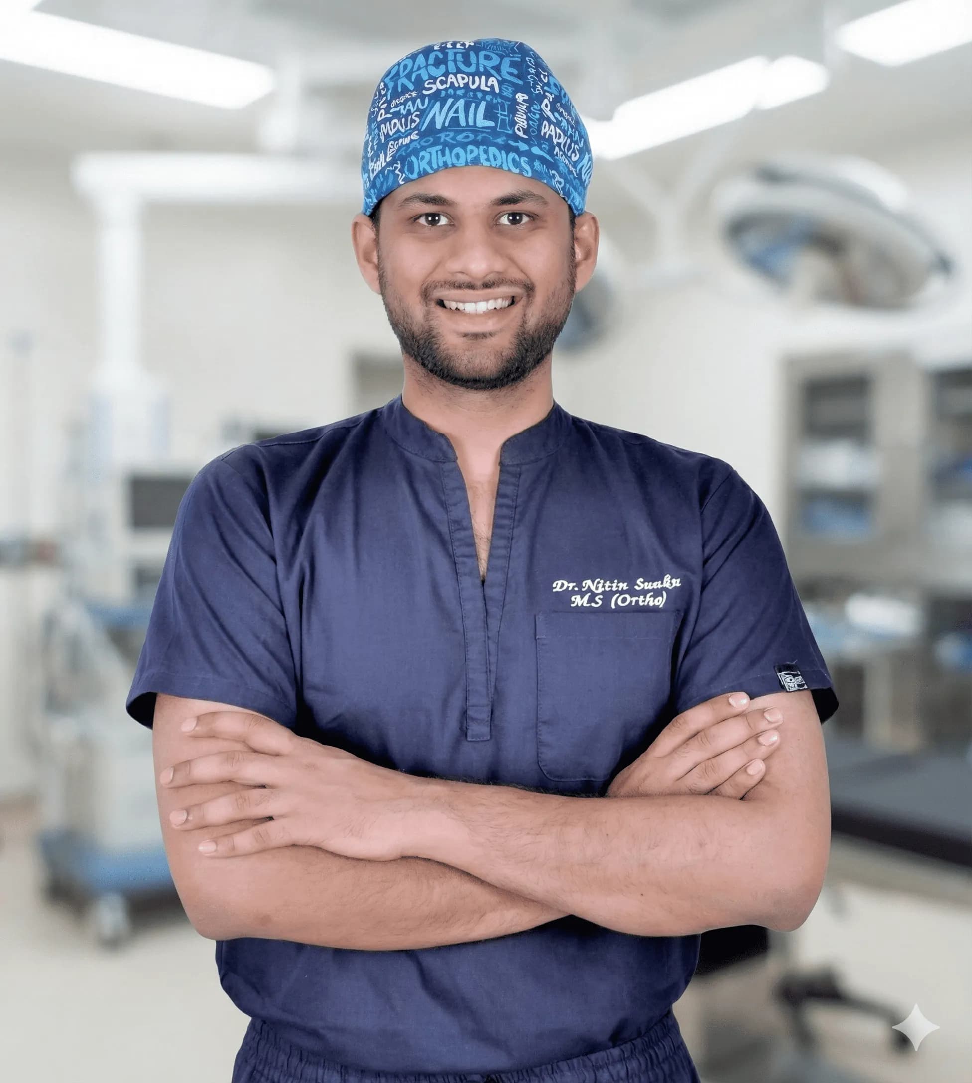
Dr. Nitin N Sunku
MBBS, MS (Orthopedics), Fellowship in Arthroscopy & Sports Medicine
Dr. Nitin N Sunku is a Consultant Orthopedic & Sports Medicine Surgeon with over 10 years of focused practice in Bengaluru. He serves as the Team Doctor for Bengaluru FC and consults at Raghava Multispeciality Hospital (Attibele) and Health Nest Hospital (HSR Layout). His clinical interests include arthroscopy, ligament & meniscus care, regenerative orthopedic medicine, ultrasound-guided injections, and joint replacement.
Experiencing symptoms?
Don't let pain hold you back. Schedule a comprehensive evaluation with Dr. Nitin today.
Book Appointment