This article is for general education and does not replace an in-person assessment, examination, or imaging. Everyone's injury pattern, medical history, and goals differ; use what you read here to prepare better questions for your doctor.
Dr. Nitin N Sunku is a consultant orthopedic and sports medicine surgeon. He sees patients at Raghava Multispeciality Hospital, Attibele, on Sarjapura–Attibele Road, and at Health Nest Hospital, HSR Layout, Bengaluru. If pain is rapidly worsening, you cannot bear weight, you develop numbness or weakness in a limb, or you have fever after an injury, seek urgent medical care. For non-emergency evaluation and individualised treatment options, book through the contact page.
Topics across this blog include knee ligament and meniscus problems, shoulder pain and instability, hip and knee arthritis, fracture recovery principles, spine symptoms when urgent causes have been excluded, running and tendon overuse issues, and what to expect from arthroscopy or joint replacement discussions. If you are comparing sources online, cross-check dates and always confirm advice with an in-person clinician.
Understand the anterior and posterior cruciate ligament (ACL and PCL), how each gets injured, how to tell them apart, and which treatments work. Expert guidance from Dr. Nitin N Sunku, orthopedic and sports medicine specialist in Bengaluru.
The knee is one of the most mechanically demanding joints in the human body. It absorbs the force of every step, twist, landing, and pivot, and it does this because of a precisely arranged system of bones, cartilage, muscles, and ligaments all working together. At the center of that system, quite literally, are two ligaments that form a cross inside the joint: the anterior cruciate ligament (ACL) and the posterior cruciate ligament (PCL).
These two structures are together responsible for the front-to-back and rotational stability of the knee. When either one is injured, the result is not just pain, it is a loss of the architectural integrity that holds the knee together during movement. Understanding what these ligaments are, how they differ, how each gets injured, and what the right treatment path looks like is one of the most useful pieces of knowledge a person can carry into a conversation with their orthopedic surgeon.
This guide covers all of it, clearly and without unnecessary complexity.
What Are the Cruciate Ligaments and Where Are They Located?
The word "cruciate" comes from the Latin word for cross. The two cruciate ligaments are named this way because they cross each other inside the knee joint, forming a visible X when the knee is opened surgically. Both ligaments connect the femur (thigh bone) to the tibia (shin bone), but they originate and insert at different points and serve complementary functions.
Together, the anterior and posterior cruciate ligaments act as the primary static stabilizers of the knee joint, meaning they do their job passively, without muscular contraction, simply by being present and intact. If one or both are torn, the knee can no longer reliably maintain its position during dynamic activities, and injury to surrounding structures like the meniscus and cartilage often follows over time.
The Anterior Cruciate Ligament (ACL)
The ACL runs from the anterior intercondylar area of the tibia, traveling upward and backward to attach on the posteromedial surface of the lateral femoral condyle. It is approximately 32 to 38 millimetres in length and 10 to 12 millimetres in width. The ACL has two functional bundles: the anteromedial bundle, which is taut in flexion, and the posterolateral bundle, which provides stability near full extension.
The primary function of the ACL is to prevent the tibia from sliding forward relative to the femur. It also resists excessive internal rotation of the tibia. Crucially, the ACL provides roughly 85 percent of the restraining force against anterior tibial displacement, which means no other structure can meaningfully compensate when the ACL is completely torn.
The ACL is the most commonly injured ligament in the knee. It does not heal on its own because of poor vascular supply and its intra-articular environment.
The Posterior Cruciate Ligament (PCL)
The PCL runs from the posterior intercondylar area of the tibia, traveling forward and upward to insert on the anterolateral surface of the medial femoral condyle. It is thicker and stronger than the ACL, with a width nearly twice that of its anterior counterpart. The PCL also has two bundles: the anterolateral bundle, which is the larger and stronger of the two, and the posteromedial bundle.
The primary function of the PCL is the opposite of the ACL: it prevents the tibia from sliding backward relative to the femur. It becomes the primary knee stabilizer when the joint is in a flexed, weight-bearing position, such as walking down a slope or descending stairs. Because of its greater thickness and strength, the PCL is less frequently injured than the ACL, but when it is damaged, the consequences for knee function and long-term joint health are significant.
How the ACL and PCL Work Together
Thinking of the ACL and PCL as a team is the most useful way to understand knee mechanics. The ACL controls forward movement of the tibia; the PCL controls backward movement of the tibia. Together, they prevent the knee from translating too far in any direction during activity.
Beyond pure translation, both ligaments also contribute to rotational stability. The ACL resists internal rotation; the PCL contributes to rotational control in the flexed knee. When the knee is functioning normally, these two ligaments work in constant coordination with the collateral ligaments (the MCL on the inner side and the LCL on the outer side) and with the quadriceps and hamstring muscle groups to produce stable, pain-free movement.
Understanding this cooperative function is why injuring one ligament often places abnormal stress on the other and on surrounding structures. An untreated ACL tear, for example, does not just create instability; over time it can lead to meniscal tears, cartilage wear, and eventually early onset arthritis in the affected knee.
How Are These Ligaments Injured?
ACL Injury: Mechanisms and Risk Factors
ACL injuries are among the most frequent serious sports injuries seen in orthopedic practice. They occur predominantly through non-contact mechanisms, meaning the player does not receive a direct blow to the knee. Instead, the ACL is typically injured when the foot is planted on the ground and the athlete rapidly changes direction, decelerates suddenly, or lands from a jump with the knee in a vulnerable position of extension or slight flexion combined with inward rotation of the lower leg.
Sports that carry particularly high ACL injury rates include football (both association and contact variants), basketball, kabaddi, volleyball, badminton, and any activity involving rapid direction changes on a firm surface.
Contact mechanisms also cause ACL tears, most commonly a blow to the outer side of the knee that forces the joint inward (valgus collapse) while the foot is planted.
Certain risk factors increase ACL injury likelihood:
- Female anatomy, due to differences in pelvis width, Q-angle, and hormonal influences on ligament laxity
- Insufficient neuromuscular control around the hip and knee
- Inadequate warm-up before explosive activity
- Playing on artificial surfaces, where traction is higher than on natural grass
- Prior ACL injury, which significantly elevates re-tear risk in the same or opposite knee
When an ACL tears, many patients report hearing or feeling a distinct pop inside the knee. Immediate swelling typically develops within a few hours, the knee feels unstable or gives way, and weight-bearing is painful and difficult.
PCL Injury: Mechanisms and Risk Factors
PCL injuries require more force than ACL tears in most cases, which explains why they occur less frequently. The classic mechanism is a direct blow to the front of a flexed knee, often called the "dashboard injury" because it is frequently seen when a patient's bent knee strikes the car dashboard in a road traffic accident. The same mechanism can occur in rugby, football, or any fall where a player lands on a flexed knee with the foot plantar-flexed.
PCL tears can also result from severe hyperflexion injuries, or from high-energy combined knee dislocations where multiple ligaments are damaged simultaneously.
Because the PCL is thicker and stronger, isolated PCL tears are relatively uncommon. More often, PCL injuries occur alongside damage to the posterolateral corner, the MCL, or even the ACL, creating what is called a multi-ligament knee injury. These combined injuries represent some of the most complex surgical challenges in orthopedic practice.
Symptoms of a PCL injury are often subtler than ACL tears. There may be mild to moderate pain and swelling, a sense of the knee feeling less secure on stairs or slopes, and aching at the back of the knee. Because the symptoms are less dramatic, PCL injuries are frequently missed or underestimated at initial assessment.
Grading Cruciate Ligament Injuries
Both ACL and PCL injuries are graded to help guide treatment decisions.
- Grade I refers to a partial tear where fibers are stretched but the ligament maintains structural continuity. The joint is stable on examination.
- Grade II refers to a partial tear with more significant fiber disruption. The ligament feels loose on stress testing but the knee has not fully lost its restraining function.
- Grade III refers to a complete tear. The ligament is fully disrupted and the knee loses its primary restraint in the affected direction. This grade typically requires surgical consideration in active patients.
- Grade IV (used specifically for PCL injuries by some classification systems) refers to a complete PCL tear combined with injury to at least one other major knee ligament, significantly increasing instability and complexity of management.
Diagnosis: How Are ACL and PCL Injuries Confirmed?
Accurate diagnosis of cruciate ligament injuries requires a combination of clinical history, physical examination, and imaging.
Clinical History
The mechanism of injury is the first and most informative clue. A non-contact pivoting injury with immediate swelling and instability strongly suggests ACL involvement. A dashboard-type impact with posterior knee pain and subtle instability on slopes raises concern for PCL damage.
Physical Examination Tests
For the ACL: The Lachman test is the most sensitive and specific clinical test. The knee is held in slight flexion while the examiner stabilizes the femur and applies a forward pull on the tibia. Abnormal forward movement (anterior tibial translation) indicates ACL insufficiency. The anterior drawer test and pivot shift test are supplementary assessments.
For the PCL: The posterior drawer test is the equivalent for PCL assessment. With the knee at 90 degrees of flexion, backward force is applied to the tibia. Abnormal posterior translation indicates PCL disruption. The posterior sag sign, where gravity alone allows the tibia to sag backward relative to the femur in a relaxed flexed position, is a useful confirmatory finding.
MRI
MRI is the gold standard imaging investigation for cruciate ligament injuries. It confirms the diagnosis, establishes grade of injury, and identifies associated pathology such as bone bruising (bone contusion), meniscal tears, or collateral ligament damage. An MRI is also essential for surgical planning, as it allows the surgeon to assess which structures need to be addressed in the operating room.
X-rays are taken to rule out bony avulsion fractures, particularly in pediatric patients where the ligament may pull away a fragment of bone rather than tearing through its substance.
Treatment Pathways: ACL vs. PCL
Treatment decisions depend on the grade of injury, the patient's age, activity level, associated injuries, and the degree of functional instability experienced in daily life.
Non-Surgical Management
Not all cruciate ligament injuries require surgery. In selected cases, non-operative management produces acceptable outcomes.
For ACL tears: Non-surgical management is most appropriate for older, less active patients, those with a partial tear showing minimal instability, or individuals whose lifestyle demands do not include cutting, pivoting, or high-demand sports. The program typically includes rest, ice, compression, and elevation in the acute phase, followed by structured physiotherapy targeting quadriceps and hamstring strengthening, proprioception training, and functional rehabilitation.
For PCL tears: Isolated Grade I and Grade II PCL tears have a better natural history than isolated ACL tears. The PCL has a somewhat better blood supply and healing potential. Many Grade I and Grade II injuries are managed conservatively with physiotherapy focused on quadriceps strengthening (which compensates for reduced PCL function by actively pulling the tibia forward) and graduated return to activity. Bracing may be used during this phase.
The non-surgical route, however, is never passive acceptance of instability. A high-quality conservative program requires consistent physiotherapy and honest re-evaluation at regular intervals to ensure the knee is stable and the patient is meeting functional milestones.
For patients exploring non-surgical options before committing to a surgical decision, the range of non-surgical knee pain treatments available at our clinic is worth understanding as part of a broader management conversation.
Surgical Management
ACL Reconstruction
ACL reconstruction is the standard surgical treatment for complete ACL tears in patients who wish to return to sports, physically demanding work, or any activity involving pivoting and cutting movements. The procedure does not repair the torn ligament directly (the ACL cannot be stitched back because it lacks healing capacity in the intra-articular environment). Instead, a graft is used to replace the ligament entirely.
Graft choices include:
- Hamstring tendon autograft (most commonly used in India): tissue harvested from the patient's own semitendinosus and gracilis tendons. Offers reliable strength, good graft biology, and a smaller donor site scar.
- Bone-patellar tendon-bone (BPTB) autograft: historically considered the gold standard for athletes due to bone-to-bone healing at tunnel interfaces. Suitable for high-demand competitive athletes.
- Peroneus longus tendon autograft: increasingly used as an alternative donor site with minimal functional loss.
- Allograft: donor tissue, used in selected revision cases or multi-ligament reconstructions.
The procedure is performed arthroscopically, meaning through small incisions with a camera and specialized instruments. The surgeon creates precisely positioned tunnels in the femur and tibia, passes the graft through these tunnels, and secures it with fixation devices that allow the graft to integrate with the bone over the following weeks and months.
Arthroscopic ACL reconstruction is minimally invasive, allows direct visualization of associated pathology such as meniscal tears, and permits early mobilization and rehabilitation.
PCL Reconstruction
Surgical reconstruction of the PCL is indicated for complete Grade III tears, multi-ligament injuries, or cases where conservative management has failed to restore functional stability. PCL reconstruction is technically more demanding than ACL reconstruction because of the proximity of critical neurovascular structures behind the knee.
The most commonly used techniques involve either a trans-tibial approach or the tibial inlay technique, where the graft is fixed directly to the posterior tibia. Graft choices are similar to ACL reconstruction, with the Achilles tendon allograft also commonly used due to its bulk for PCL reconstruction.
When the PCL injury is part of a multi-ligament knee injury, surgical staging and careful planning of which structures to address first (and in what order) becomes critical to achieving a stable outcome without compromising any single reconstruction.
For patients with combined ligament injuries or knee instability that has not been adequately addressed, the ACL care service page provides more detail on how these cases are evaluated and managed in our clinic.
Recovery After Cruciate Ligament Surgery
Rehabilitation after ACL or PCL reconstruction is a structured, phased process. Recovery timelines are not arbitrary; they reflect the biology of graft incorporation and neuromuscular re-education, both of which cannot be rushed without increasing failure risk.
- Phase One (Weeks 0 to 2): The focus is on controlling swelling, reducing pain, and achieving early range of motion. Walking with crutches begins immediately. Ice, elevation, and anti-inflammatory management are standard.
- Phase Two (Weeks 2 to 6): Quadriceps activation, gentle strengthening, proprioception training, and progressive weight-bearing. The goal is to walk without crutches and regain 0 to 90 degrees of flexion.
- Phase Three (Months 2 to 4): More aggressive strengthening, balance and coordination drills, and sport-specific movement patterns are introduced cautiously.
- Phase Four (Months 4 to 6): Running, agility training, and sport-specific drills for athletes. Return-to-sport testing is performed before clearance is given.
Return to competitive sport typically occurs at nine to twelve months post-surgery for ACL reconstruction, with growing evidence that returning before nine months significantly increases re-tear risk. PCL reconstruction has a broadly similar timeline, though multi-ligament reconstructions may require longer rehabilitation.
Understanding what post-surgical warning signs to watch for during recovery is equally important. Patients who want to know what to monitor during their healing process can find a useful overview in our blog on bad signs after ACL surgery that applies equally to anyone recovering from cruciate ligament procedures.
Can Cruciate Ligament Injuries Be Prevented?
No injury prevention program eliminates risk entirely, but structured neuromuscular training programs have demonstrated meaningful reductions in ACL injury rates in multiple clinical trials. These programs target the landing mechanics, deceleration patterns, and lower limb muscle activation sequences that create vulnerability when they are poorly controlled.
Key prevention elements include:
- Single-leg landing and balance exercises to develop unilateral stability
- Hip abductor and external rotator strengthening to prevent knee valgus on landing
- Plyometric training with attention to landing technique (soft landings with hip and knee flexion rather than stiff-legged contact)
- Hamstring strengthening, particularly eccentric work (Nordic hamstring curls and similar exercises)
- Education about high-risk movement patterns specific to the player's sport
For athletes recovering from one cruciate ligament injury and returning to sport, structured prevention work is especially important because re-injury rates in the previously affected knee, and in the contralateral knee, are meaningfully elevated for at least two years after reconstruction.
The broader picture of how sports injuries are managed and how return-to-play decisions are made safely is covered in more depth in our overview of advanced sports injuries treatment.
Consulting a Specialist in Bengaluru for Cruciate Ligament Injuries
Cruciate ligament injuries are not emergencies in the same way a fracture is, but they are not conditions to monitor passively either. Delayed diagnosis and inadequate rehabilitation increase the risk of secondary damage to the meniscus and articular cartilage, which can accelerate the development of knee arthritis and reduce the long-term success of any future surgical intervention.
If you have experienced a knee injury with swelling, instability, or a feeling that the knee is giving way, an early assessment by a fellowship-trained orthopedic and sports medicine specialist is the most important step you can take.
Dr. Nitin N Sunku is a fellowship-trained orthopedic surgeon specializing in arthroscopy and sports medicine. He serves as the Team Doctor for Bengaluru FC and has managed cruciate ligament injuries across a full spectrum of patients, from professional athletes to working adults and adolescents participating in school sports. His approach begins with a thorough clinical examination and MRI review, followed by an honest conversation about whether surgery is necessary, what the realistic recovery timeline looks like, and what the risks of both operative and non-operative management are.
Consultations are available at Raghava Multispeciality Hospital, Attibele (serving patients from Electronic City, Chandapura, Anekal, Jigani, Bommasandra, and Sarjapura) and at Health Nest Hospital, HSR Layout (accessible to patients from Koramangala, BTM Layout, Bellandur, and surrounding south Bengaluru localities).
You can explore the range of knee and sports medicine services available by visiting the services overview or by calling +91-9980031006 to schedule an appointment.
Resource References
These resources provide peer-reviewed and clinically authoritative background on cruciate ligament anatomy and injury management:
- American Academy of Orthopaedic Surgeons (AAOS) — OrthoInfo on PCL injuries: orthoinfo.aaos.org
- StatPearls (NCBI) — Knee Anterior Cruciate Ligament Anatomy: ncbi.nlm.nih.gov/books/NBK559233
- StatPearls (NCBI) — Knee Posterior Cruciate Ligament Anatomy: ncbi.nlm.nih.gov/books/NBK535416
- Cleveland Clinic — Knee Ligaments: my.clevelandclinic.org/health/body/21596-knee-ligaments
Frequently Asked Questions About the Anterior and Posterior Cruciate Ligament
Q1: What is the difference between the anterior and posterior cruciate ligament?
The anterior cruciate ligament (ACL) prevents the tibia from sliding forward relative to the femur and resists inward rotation of the lower leg. The posterior cruciate ligament (PCL) prevents the tibia from sliding backward and provides stability when the knee is bent and bearing weight. Together they form a cross inside the joint, giving the knee front-to-back and rotational control.
Q2: Which is worse to injure: the ACL or the PCL?
Both ligaments are important and both injuries have significant consequences if untreated. However, ACL tears are generally considered more functionally disabling in the short term because of the greater instability they cause during everyday activities and sports. PCL injuries are often subtler in their immediate presentation but can cause serious long-term problems including early arthritis if left unmanaged.
Q3: Can cruciate ligament injuries heal without surgery?
Grade I and Grade II PCL tears have reasonable healing potential with non-surgical management. Complete ACL tears (Grade III) do not heal on their own because the intra-articular environment prevents natural tissue repair. Non-surgical management of a complete ACL tear can allow return to low-demand activities but is generally not appropriate for patients who want to return to pivoting or cutting sports.
Q4: How is an ACL or PCL injury diagnosed?
Diagnosis involves a combination of clinical history, physical examination tests (such as the Lachman test for the ACL and the posterior drawer test for the PCL), and MRI imaging. MRI is the most accurate investigation, confirming the degree of injury and identifying any associated damage to the meniscus, cartilage, or other ligaments.
Q5: How long does recovery take after ACL or PCL reconstruction?
Most patients return to daily activities and light work within six to eight weeks of surgery. Return to sports, particularly cutting and pivoting sports, typically requires nine to twelve months after ACL reconstruction. Multi-ligament reconstructions involving both the ACL and PCL may require longer rehabilitation, sometimes up to twelve to eighteen months for full return to competitive sport.
Q6: Is ACL reconstruction performed arthroscopically?
Yes. Arthroscopic ACL reconstruction is the standard surgical technique. It involves small incisions through which a camera and specialized instruments are introduced. This minimally invasive approach reduces post-operative pain, lowers infection risk, allows earlier mobilization, and provides the surgeon with excellent visualization of associated pathology inside the joint.
Q7: Can both the ACL and PCL be injured at the same time?
Yes. Combined ACL and PCL injuries typically occur in high-energy trauma such as road traffic accidents or contact sports collisions involving knee dislocation. These multi-ligament injuries are among the most complex cases in orthopedic surgery and require careful pre-operative planning. The majority require surgical reconstruction, often staged across more than one procedure.
Q8: What happens if a cruciate ligament injury is left untreated?
Leaving a significant cruciate ligament injury untreated leads to ongoing knee instability. Over time, this instability causes increased stress on the menisci and articular cartilage, accelerating their damage. Patients who do not address an ACL or PCL injury appropriately are at significantly higher risk of developing early knee osteoarthritis compared to those who receive timely, appropriate treatment.
Q9: What is the best graft for ACL reconstruction?
There is no single universally best graft. The choice depends on factors including the patient's age, sport, activity demands, and the surgeon's experience. Hamstring tendon autograft is the most commonly used graft in India and is appropriate for most patients. Bone-patellar tendon-bone autograft is preferred by some surgeons for high-demand athletes. The right choice is a shared decision made after examining the patient's anatomy and discussing their functional goals.
Q10: Where can I get cruciate ligament assessment and treatment in Bengaluru?
Dr. Nitin N Sunku sees patients with ACL and PCL injuries at Raghava Multispeciality Hospital, Attibele and Health Nest Hospital, HSR Layout in Bengaluru. To book a consultation, visit the contact page or call +91-9980031006.
This article is for educational purposes and should not substitute for a clinical evaluation. If you have acute knee trauma, inability to bear weight, significant swelling, or a feeling that the knee is dislocated, seek emergency care immediately. For all other knee concerns, early specialist assessment is the recommended first step. Dr. Nitin N Sunku — Orthopedic and Sports Medicine Specialist | Attibele and HSR Layout, Bengaluru, Karnataka.
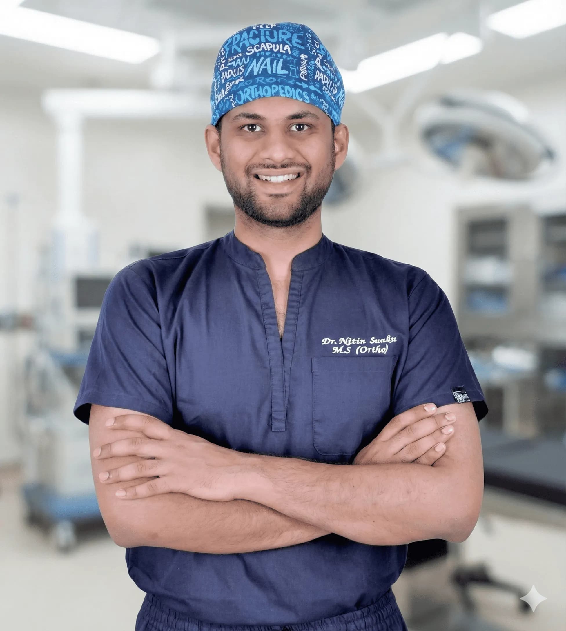
About the Author
Dr. Nitin N Sunku
MBBS, MS (Orthopedics), Fellowship in Arthroscopy & Sports Medicine
Dr. Nitin N Sunku is a Consultant Orthopedic & Sports Medicine Surgeon with over 10 years of focused practice in Bengaluru. He serves as the Team Doctor for Bengaluru FC and consults at Raghava Multispeciality Hospital (Attibele) and Health Nest Hospital (HSR Layout). His clinical interests include arthroscopy, ligament & meniscus care, regenerative orthopedic medicine, ultrasound-guided injections, and joint replacement.
Experiencing symptoms?
Don't let pain hold you back. Schedule a comprehensive evaluation with Dr. Nitin today.
Book Appointment